Research Overview
Highly conserved mismatch repair (MMR) systems have been identified in organisms ranging from bacteria to humans that recognize and repair base pair and small insertion/deletion mismatches that arise as the result of DNA replication errors, DNA damage, and genetic recombination. These systems are thought to improve the fidelity of DNA replication by about three orders of magnitude. In humans, mutations in MMR genes have been correlated to both an increased mutation rate and a predisposition to a hereditary form of colorectal cancer (HNPCC). The goal of our laboratory is to understand how MMR proteins interact to prevent replication errors and to modulate genetic recombination. We are addressing these questions in the model organism baker's yeast (Saccharomyces cerevisiae) using a combination of genetic and biochemical approaches.
In eukaryotes the MSH (MutS homolog) and MLH (Mut L homolog) family proteins play key roles in early steps in MMR. The MSH proteins recognize DNA mismatches and the MLH proteins interact with the MSH-mismatch DNA complex to signal downstream repair factors such as helicases and exonucleases. MMR factors also excise mismatches formed in heteroduplex DNA, prevent recombination between divergent DNA sequences, and process recombination intermediates containing non-homologous single-stranded ends. In higher eukaryotes these factors also act as DNA damage sensors to regulate cell cycle progression and entry into apoptotic pathways.
The mechanisms by which MMR proteins identify mismatches and signal downstream factors during DNA replication and recombination are not well understood. In addition, the role of genetic background in determining the effect of MMR mutations with respect to the severity of disease has not been explored in depth. We use genetic, biochemical, single-molecule, and population genetic approaches to examine interactions between the MSH-MLH mismatch recognition complex and other DNA repair, recombination, and replication factors. Current projects in the lab are described below.
Project 1. Single-molecule approaches to test mechanistic models for MMR
We are using single-molecule approaches to measure the specificity of MSH proteins for mismatch DNA and to track MSH2-MSH6 on DNA in real time. Through unzipping force analysis (in collaboration with Michelle Wang, Dept. of Physics, Cornell) we detected high affinity mismatch binding and sliding clamp modes for MSH2-MSH6 (Jiang et al. Mol. Cell 20, 771). In collaboration with the Greene lab at Columbia University, we are utilizing total internal reflection fluorescence microscopy to monitor movement of MSH proteins along DNA (Gorman et al. Mol. Cell 28, 359). These studies are aimed at understanding how MSH proteins can identify DNA mismatches with high specificity. These studies will take advantage of a large number of MMR mutants generated previously in the lab and are aimed at distinguishing between competing models for how MSH and MLH proteins signal downstream steps in MMR.

MMR improves the fidelity of DNA replication in E. coli by about 1000 fold by excising DNA mismatches in the newly replicated strand that arise from polymerase misincorporation and slippage. In E. coli MMR is initiated by MutS binding to mismatch DNA. ATP-dependent recruitment of MutL to mismatch-MutS complexes is important for transmitting the mismatch recognition signal to key downstream repair factors including MutH, an endonuclease that nicks the newly replicated DNA strand at unmethylated GATC sites. The net result of these actions is the unwinding of DNA by UvrD helicase from the nick towards the mismatch site. This is followed by excision of the unwound newly synthesized strand. The resulting gap is repaired by DNA polymerase III and DNA ligase in conjunction with single-strand DNA binding protein.

Duplex DNA molecules (1.1 kb) containing single DNA mismatches are unwound in the presence or absence of the MSH2-MSH6 mismatch binding complex. Unwinding is accomplished by anchoring one end of a duplex DNA to a microscope coverslip binding chamber through a digoxigenin/anti-digoxigenin linkage. This substrate contained a biotinylated base adjacent to a nick located in the middle of duplex DNA. This base is attached to a streptavidin-coated microsphere held in a feedback-enhanced optical trap. DNA becomes unzipped as the coverslip is moved away from the optical trap, simultaneously recording the coverslip position. The unzipping force yields a force curve that can detect the presence of a protein bound to a specific site on a DNA substrate due to the transient obstruction of the unzipping fork at the protein-DNA interface. The obstruction results in an increase in the unzipping force required to unwind DNA followed by a sudden drop in the force once the protein-DNA complex has been disrupted. We found that unzipping force analysis could detect the presence of single mismatches in duplex DNA at 2-3 bp resolution as measured by a decrease in unzipping force centered at the mismatch site.

A model showing how the rejection of recombination intermediates containing mismatch DNA (heteroduplex rejection) and non-homologous tail removal act to modulate genetic recombination that occurs through single-strand annealing. In the left branch, MSH2-MSH6 recognizes DNA mismatches that form in the single-strand annealing intermediate and recruits the SGS1 helicase to unwind the annealed region. In the absence of heteroduplex rejection, the SSA intermediate is repaired through non-homologous tail removal, which is thought to involve MSH2-MSH3 binding to branched structures, followed by RAD1-RAD10 cleavage of the single-strand tail, and DNA synthesis and ligation steps (right branch). See Goldfarb and Alani, Genetics 169, 563-574.
Project 2. Understanding how MMR proteins act to reject recombination between divergent DNA sequences
Recombination between divergent sequences in the genome can lead to chromosomal translocations, deletions, or inversions which in higher eukaryotes are thought to contribute to tumor formation. MMR proteins act to prevent such recombination. We are interested in understanding this process at the molecular level using a simple recombination assay developed by Jim Haber's laboratory at Brandeis University. In this system a double-strand break is induced between two regions of the chromosome that are either homologous or show 3% sequence divergence. The break is then processed by exonucleases to generate single-stranded DNA tails which then pair and anneal at homologous sequences. The annealed sequences are then sealed after steps that include non-homologous tail removal, DNA synthesis, and ligation. In this system the mismatch repair protein MSH6 and the SGS1 helicase play equally important roles in preventing recombination between divergent DNA sequences, presumably during the single-strand annealing process (Sugawara et al. PNAS 101:9315). In contrast, other mismatch repair factors such as MLH1, PMS1 and EXO1, were shown to not be required, or to play only minimal roles. These findings provide a nice illustration for how subsets of mismatch repair proteins collaborate with different DNA repair factors to maintain genome stability.
We tested mutations that disrupt SGS1 helicase activity and MSH2-MSH6 mismatch recognition and ATP binding and hydrolysis activities for their effect on preventing recombination between divergent DNA sequences (Goldfarb and Alani, Genetics 169, 563-574). These studies led to a model in which the MSH proteins act in concert with the SGS1 helicase to unwind DNA recombination intermediates containing mismatches. We are testing this model in vitro using wild-type and mutant forms of MSH2-MSH6 and SGS1 and DNA substrates predicted to form during single-strand annealing between divergent DNA sequences.
Project 3. Identification of genetic incompatibilities that result in defects in MMR
The MLH1-PMS1 heterodimer is the major MutL homolog complex acting in mismatch repair in baker’s yeast. Using a highly sensitive mutator assay, we observed that yeast strains bearing the S288c MLH1 allele and the SK1 PMS1 allele displayed elevated mutation rates that conferred a long-term fitness cost in diploid strains. The S288c and SK1 strains display ~0.7% DNA sequence divergence. Dissection of this defect revealed that only a single amino acid polymorphism in each gene accounts for this defect. Were these strains to cross in natural populations, segregation of these alleles would generate a mutator phenotype that while potentially transiently adaptive, would ultimately be selected against due to the accumulation of deleterious mutations. Such fitness “incompatibilities” could potentially contribute to reproductive isolation among geographically dispersed yeast (Heck et al. PNAS 103:3256, Demogines et al. PLoS Genetics 4:e1000103). We are testing this hypothesis using a population genetic approach in collaboration with the Aquadro lab at Cornell.
The segregational mutator phenotype described above may underlie some cases of a HNPCC, as well as some sporadic cancers. Many cases of familial colorectal cancer do not display the clear dominant inheritance of typical HNPCC families, and there are numerous MMR gene variants whose associations to cancer predisposition are unclear (http://www.nfdht.nl). The negative epistatic interaction observed between yeast MMR gene variants suggests a mechanism to explain the genetic basis of some HNPCC-like cancers displaying atypical inheritance. We are testing this idea by making mutations in the yeast MLH1 gene that correspond to those found in HNPCC databases and then testing the effect of these mutations in the SK1 and S288c yeast strains (Wanat et al. Hum. Mol. Genet. 16:445).

Likely reconstruction of events resulting in the observed defect in MMR in S. cerevisiae gene pool. 65 S. cerevisiae strains are grouped according to their amino acid residues 761 (G or D) in MLH1 and 818/822 (R or K) in PMS1. Bold arrows indicate transitions between genotypes resulting from single mutational events. The relative mutation rates of genotypes, based on Lys+ reversion experiments using S288c and SK1 strains (1X = wild-type S288c), are shown. Genetic exchange between D-R and G-K strains would generate a mutator combination (D-K) at a 25% frequency (indicated by thick dotted arrows).

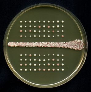
Losing track of time through yeast tetrad analysis.
Project 4. Roles for MMR proteins in meiotic crossing over
In most eukaryotic organisms, the correct segregation of chromosomes at the Meiosis I division requires crossing over (reciprocal exchange) between homologs. Crossing over is highly regulated during meiosis with respect to the number of events, their placement, and timing. In S. cerevisiae, two crossovers rarely occur within the same genetic interval (crossover interference), and every chromosome, regardless of size, receives at least one reciprocal exchange. In humans, defects at the Meiosis I division can lead to miscarriage and a variety of chromosome aneuploidy syndromes including Down’s, Turner’s, and Kleinfelter's. We built a S. cerevisiae strain and designed software to analyze meiotic crossing over at consecutive genetic intervals (Argueso et al. Genetics 168:1805). In addition we are working with strains that allow us to score crossover events on chromosomes that have suffered a MI non-disjunction event. These tools, in combination with physical and cell biological approaches, are aimed at understanding why mutations in subsets of MSH and MLH homologs (e.g. mlh1Δ, mlh3Δ, msh4Δ, msh5Δ) as well as mutations in genes whose products act in telomere-led chromosome dynamics (csm4Δ, ndj1Δ), confer high levels of Meiosis I non-disjunction (Nishant et al. Genetics 179:747, Wanat et al. PLoS Genetics 4 e1000188; Zanders and Alani PLoS Genetics 5, e1000571).